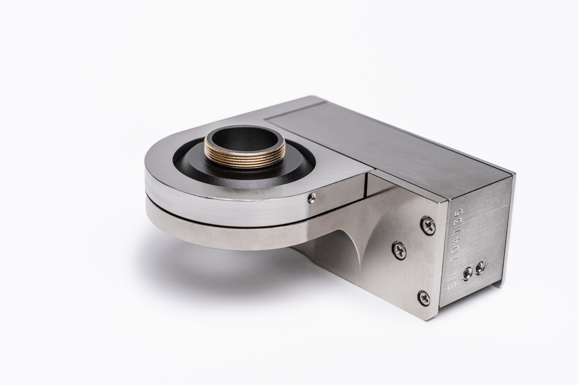
NanoScan OP800 Piezo Objective Positioner/Scanner With 800 µm Range
The NanoScan OP800 piezo-driven objective positioner is compatible with most commercial and customized microscopes. The stage features an M32 objective thread as standard with a wide range of adapters available.
Settings for inverted or upright operation and load capacity are set during manufacture and can be further tuned in the field to optimize for specific applications.
The OP800 provides the fastest step and settle time of any piezo-driven objective positioner with a range of up to 800 µm. It features capacitive feedback sensors that ensure exceptional repeatability and resolution.
With its long travel range, it is especially suitable for whole organism, lightsheet, and multiphoton microscopy.
0-500 g load as standard: a high load version (up to 1000 g) is available.
“We have recently added the Prior NanoScan OP800 to our imaging setup. We have had success using this device. It was easy to install onto our microscope nosepiece using brass adapter rings and we have easy control using the included software. For our imaging, our objective is to capture neuronal fluorescence that is distributed over a curved surface. On its own, our objective only allows us to visualize a small portion of this surface corresponding to a narrow focal plane. The NanoScan OP800 has enabled us to double the effective measurement field. We have programmed the device to cover a distance of 300 μm in 100 μm steps during our acquisition window of 200 ms. The device can step through these planes almost instantaneously and we observe no obvious movement artifact. We find a decrease in resolution that is expected from acquiring in multiple planes. Post-experiment image processing, reveals that this diminished resolution does not interfere with our downstream analysis. Importantly, we only find that we gain additional fluorescent signal in the previously out-of-focus planes without any information loss from the original, narrower focal field. We have attached processed images from our recording sessions of equivalent neuronal fluorescence acquired with and without the NanoScan OP800 to demonstrate the difference in our data.”
Joshua J. Emrick, DDS, PhD at The University of Michigan School of Dentistry
Image acquired without NanoScan OP800
Image acquired with NanoScan OP800
Images are of a GCaMP6f fluorescence in the trigeminal ganglion Courtesy of Akash R. Gandhi, Elizabeth A. Ronan, PhD, and Joshua J. Emrick, DDS, PhD, Department of Biologic and Materials Sciences & Prosthodontics, University of Michigan School of Dentistry, Ann Arbor, Michigan.
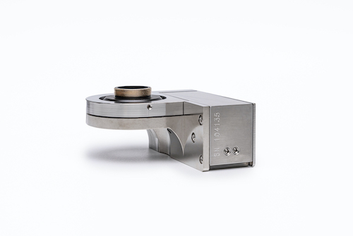
800μm closed loop travel range (950μm open loop range)
Capacitive positioning sensors give sub nanometer positioning resolution and repeatability
Made from stainless steel providing greater mechanical stiffness (faster) and temperature stability (lowest drift).
The stage is a flexure guided system. The friction free flexures are designed to provide high stiffness and to minimize off axis motions giving high repeatability and faster cycle times.
Options for upright and inverted microscopy applications.
Connectors with built in stage calibration provide plug and play electronics which can be interchanged, minimizing system down times.
Rapid settling times even with large objective loads. Tested to function for greater than 10 million full range cycles.
Please provide as much information to help us assist you. A Queensgate representative will respond to your request.
If you are an existing customer with a technical support request, please use the customer support form